Stem Cells
The Origin of Breast Cancer | Drew Berry, Etsuko Uno
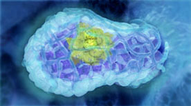
This animation visualises research published in Nature Medicine (Vol 15, Issue 8, 2009) by the laboratory of Jane Visvader and Geoffrey Lindeman. The mammary gland is comprised of three main cell types; alveolar, ductal and myoepithelial cells. Breast stem cells can develop into any of the three cell types through a series of intermediate cell stages. One intermediate is the luminal progenitor cell, which develops into either alveolar or ductal cells. The paper describes how an aberrant form of a luminal progenitor cell is involved in the development of some forms of breast cancer.
» View the animationThe Control of Breast Stem Cells | Drew Berry, Etsuko Uno
This animation illustrates how breast stem cells respond to steroid hormone despite the cells not having any steroid receptors. The animation illustrates the research published in Nature (Vol 465, Issue 7299, 2010) by the laboratory of Jane Visvader and Geoffrey Lindeman.
» View the animationBreast Stem Cells | Drew Berry, Etsuko Uno
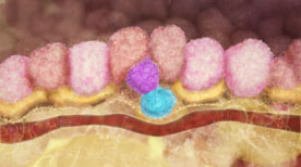
An overview of the human mammary gland with a focus on the role of breast stem cells during pregnancy. The primary function of the mammary gland is to produce milk to nourish young offspring. The mammary gland is comprised of three main cell types; alveolar, ductal and myoepithelial cells. During pregnancy, the mammary gland increases in size due to the action of breast stem cells, which can mature into any of the three mammary gland cell types.
» View the animationStem Cell Introduction | Arkitek Studios
A series of animations with audio and text commentary that clearly explain the basics of stem cell biology (including their unique characteristics, pluripotency in the early embryo, presence in adult tissues and embryonic stem cells in culture).
» View the animationIntestinal Crypt Stem Cells – A Clonal Conveyor Belt | Digizyme, Eric Keller
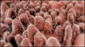
This animation, created for Hans Clevers’ lab, shows how the entire surface of the intestine is populated via a “clonal conveyor belt” mechanism. Daughter cells born from stem cells located at the base of the crypts travel up and differentiate, thereby pushing existing cells up towards the villus tip (the oldest cells are jetisoned via apoptosis at the villus tip). Adenoma formation is also shown.
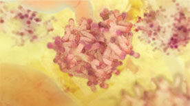
» View the animationCSF Receptor | Drew Berry
A molecular view of the surface of a stem cell highlighting the binding of G-CSF by its receptor, dimerization, signal transduction and the resulting effect on cell division and growth.
» View the animation at WEHI