Stylus Visuals
Viral DNA Packaging – Part I | Eric Keller, Stylus Visuals
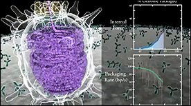
This 2-part Maya animation depicts the process of nucleic acid packing/assembly into the viral capsid. Part I shows the process simultaneous with the measured kinetics of packing and force (displayed on the right).
» View the animationViral DNA Packaging – Part II | Eric Keller, Stylus Visuals
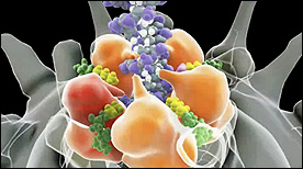
This 2-part Maya animation depicts the process of nucleic acid packing/assembly into the viral capsid. Part II focuses on the molecular machinery responsible for pulling the nucleic aacid strand inside the capsid.
» View the animationCapsid Molecular Dynamics Simulation | Geordie Martinez, Stylus Visuals
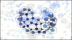
A Maya-rendered visualization of a VMD molecular dynamics simulation. Created for David Chandler’s lab at UC Berkeley, this movie depicts the physics of viral capsid formation while summarizing some of the technical steps involved in its creation.
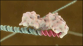
» View the animationSignal Recognition Particle | Eric Keller, Steve Davy, Stylus Visuals
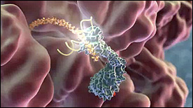
This Maya animation depicts the process by which the translating ribosome is halted by the signal recognition particle (SRP). The ribosome is subsequently brought to the membrane and docked with a channel to translocate the nascent polypeptide chain.
» View the animation at BloopatoneIRES | Stylus Visuals
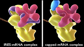
This animation compares the structure of ribosome complexes in either IRES-mRNA (Internal RIbosome Entry Sequence) or capped-mRNA conformations.
» View the animationDicer | Geordie Martinez, Steve Davy, Stylus Visuals
This Maya animation shows cleavage of double-stranded RNA into short RNA fragements by the Dicer ribonuclease.
» View the animationMicrotubules: Structure, Function & Dynamics | Geordie Martinez, Steve Davy, Stylus Visuals
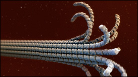
This Maya animation depicts the dynamic self-assembly and dissassembly processes of microtubules. The animation incoporates atomic resolution structural information for tubulin (as it undergoes a GTP vs GDP-induced conformational change), as well as cryoEM data for ‘protofilament peels’ and ‘helical ribbons’ from the Nogales lab.
» View the animation