Drew Berry
The Lifecycle of Malaria (Part 1) | Drew Berry
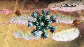
This animation represents part-1 of a 2-part series depicting the events of the malaria parasite lifecycle.
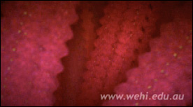
The parasite is shown entering the human host following a mosquito bite and we follow its progression initially to the liver and subsequently targeting erythrocytes on a large scale.
» View the animationThe Lifecycle of Malaria (Part 2) | Drew Berry
Part 2 depicts events in the mosquito host. The malaria parasite is shown reproducing in the mosquito’s stomach followed by the development of cysts and infection of the salivary glands.
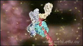
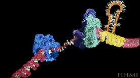
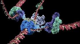
» View the animationTranslation | Drew Berry
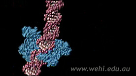
Part 2 in Drew Berry’s “Central Dogma” animations – the mRNA (yellow) is decoded inside the ribosome (purple and light blue) and translated into a chain of amino acids (red) as aminoacyl-tRNAs (green) deliver each amino-acid cargo (red/pink tip) to the ribosome.
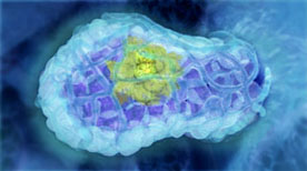
» View the animation at WEHIGolgi / ER Visualization | Drew Berry
A visualization of a cell’s cytosplasm derived from electron tomography data from Brad Marsh’s laboratory. The different components – nucleus, microtubules, mitochondria, ribosomes, smooth ER, rough ER, Golgi – are highlighted in separate ‘passes’ and then overaid as one. A great reminder of how crowded cellular interiors are!
» View the animationDNA Central Dogma Part 1 – Transcription | Drew Berry
Transcription factors assemble at a DNA promoter region found at the start of a gene. Promoter regions are characterised by the DNA’s base sequence, which contains the repetition TATATA É and for this reason is known as the “TATA box”.
» View the animation at WEHIThe Origin of Breast Cancer | Drew Berry, Etsuko Uno
This animation visualises research published in Nature Medicine (Vol 15, Issue 8, 2009) by the laboratory of Jane Visvader and Geoffrey Lindeman. The mammary gland is comprised of three main cell types; alveolar, ductal and myoepithelial cells. Breast stem cells can develop into any of the three cell types through a series of intermediate cell stages. One intermediate is the luminal progenitor cell, which develops into either alveolar or ductal cells. The paper describes how an aberrant form of a luminal progenitor cell is involved in the development of some forms of breast cancer.
» View the animationThe Control of Breast Stem Cells | Drew Berry, Etsuko Uno
This animation illustrates how breast stem cells respond to steroid hormone despite the cells not having any steroid receptors. The animation illustrates the research published in Nature (Vol 465, Issue 7299, 2010) by the laboratory of Jane Visvader and Geoffrey Lindeman.
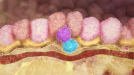
» View the animationBreast Stem Cells | Drew Berry, Etsuko Uno
An overview of the human mammary gland with a focus on the role of breast stem cells during pregnancy. The primary function of the mammary gland is to produce milk to nourish young offspring. The mammary gland is comprised of three main cell types; alveolar, ductal and myoepithelial cells. During pregnancy, the mammary gland increases in size due to the action of breast stem cells, which can mature into any of the three mammary gland cell types.
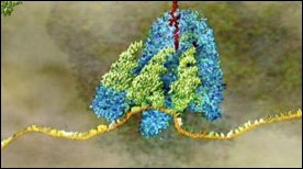
» View the animationCSF Receptor | Drew Berry
A molecular view of the surface of a stem cell highlighting the binding of G-CSF by its receptor, dimerization, signal transduction and the resulting effect on cell division and growth.
» View the animation at WEHITri Nucleotide Repeat | Biointeractive.org, Drew Berry, HHMI
This animation shows how a tri-nucletoide repeat can cause the DNA polymerase to ‘slip’ and incorporate additional nucleotides during the replication process.
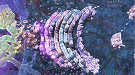
» View the animation at HHMIReplication | Drew Berry
Still one of the more complex and beautiful molecular animations ever made, this movie shows the components and dynamic processes involved in the replication of both the leading and lagging strands of DNA.
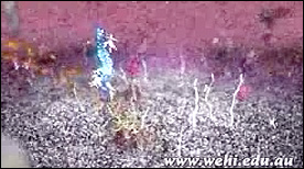
» View the animation at WEHIThe Whole Brain Catalog | Drew Berry
A visualization of the possibilities of the Whole Brain Catalog (http://wholebraincatalog.org), an open source, multi-scale virtual catalog of the mouse brain.
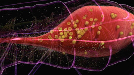
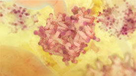
» View the animationSickle Cell Hemoglobin | Drew Berry
This animation depicts hemoglobin molecules binding to oxygen. The mutant form of hemoglobin is also shown and results in the assembly of the long stiff protein fibers characteristic of the disease sickle cell anemia.
» View the animation at WEHIRestriction Endonuclease Digestion & Ligation | Drew Berry
This animation depicts the proces of DNA recombination. The DNA plasmid is first digested with the restriction endonuclease enzyme ecoRI. Then, a piece of DNA encoding a gene is inserted into the plasmid by DNA ligase.
» View the animation at WEHIDNA structure | Drew Berry
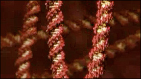
A series of short animations highlighting the structureand flexibility of the DNA double-helix.
» View the animation at WEHIChromatin | Drew Berry
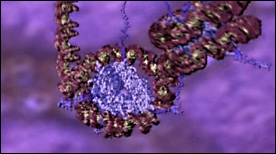
This animation shows the different levels of chromatin packing – starting with wrapping of DNA around histone octamers and nucleosome assembly, all the way to chromosome condensation during mitosis.
» View the animation at WEHIApoptosis | Drew Berry
This stunning Maya animation covers the death receptor signaling pathway that originates with binding of the Fas/TNF family of ligands, triggering of the caspase cascade, cytochrome C release from the mitochondria, apoptosome activation, and ensuing signal amplification.
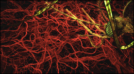
» View the animationAngiogenesis | Drew Berry
This animation shows the process by which tumors recruit new blood vessels thereby facilitating the metastatic behavior of stray cells that enter the circulation.
» View the animation